White Spots in Beef Liver Safe to Eat
Author: Mark White BVSc LLB DPM MRCVS
Reviewed: Mark White BVSc LLB DPM MRCVS 2016
Published: 2007
There are a limited number of worm parasites that affect the pig in the UK. The most prevalent of these is Ascaris suum, most commonly associated with milk spot liver. Whilst severe infestations are generally only associated with the poorest levels of hygiene, modest levels of worms are present in many herds and can have a significant effect on growth and feed efficiency.
Over the last 11years producers have had access to abattoir reports (generated under the British Pig Health Scheme) clarifying questions regarding milk spot liver condemnation and the need for control of the disease on farm.
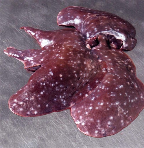
Fig1 Extensive milk spot in a single liver. (Photo courtesy of British Pig Health Scheme)

The different type of lesions that migrating Ascariasis suum larvae. (Photo courtesy of British Pig Health Scheme) Lymphonodular lesions in the centre (The 2 linear marks are unrelated hepatic capsular scars)

Fig 2b Dispersed lymphoid infiltration
The lesions are found within the tissue of the liver - not just on the surface - and can take the form of discrete lymphonodular accumulations (Fig 2a) or more commonly dispersed lymphoid accumulations - hence the term milk spot. Very early lesions may contain a small haemorrhagic centre where the larva has penetrated the capsule. Migrating larvae do not induce the formation of scar tissue (fibrosis) which explains why they resolve in a matter of weeks - generally fibrous tissue once formed is permanent.

Fig 3 Intestinal section completely blocked with Ascaris suum worms - an extremely rare occurrence in growing pigs
Like all worm parasites, there is a complex life cycle with Ascaris suum. The adult worm - which can be up to 40cm long - lives in the small intestine of the pigs; in sows there may only be a few of these worms present. Each adult produces huge quantities of eggs intermittently and so examination of faeces for worm eggs can be an unreliable method of diagnosis.
The specific feature of the eggs of most significance is that they are covered with a sticky protective coat, which means that the egg will survive many years outside the body of the host. Its sticky nature makes it difficult to wash away by cleaning and allows very easy spread between units on animal and mechanical carriers. Birds are probably quite significant in the spread of the eggs. The protective coat also renders the egg very resistant to drying and disinfection. The only reliable methods of destroying the eggs are with fire (flame gun) or caustic soda. Oocide (Antec) may also be effective, although the nature of this product is such that it is difficult to use in the types of buildings where the eggs build up e.g. dry sow housing and grower sheds.
In the environment, the eggs undergo a maturation phase, which occurs more rapidly in higher temperatures but will take at least 2 weeks. This phase leads to the hatching of a larva, which will then be ingested by the pig. There is reason to believe that the sucking pig receiving milk is resistant to these larvae but the eggs can easily be picked up in the farrowing area, stuck to the body and then mature and infect after weaning.
Once the larva has been swallowed, it will begin one of its most destructive phases. The larva penetrates the wall of the intestine and migrate around the body specifically first to the liver and then to the lungs, all the while continually maturing. Eventually the larvae will be coughed up, re-swallowed and re-enter the gut to mature to adult worms. The whole life cycle will take a minimum of 8 weeks.
Due to the effects of temperature on larval development outside the body there is a marked seasonality in the incidence of milk spot with levels typically rising in late summer and autumn. This was particularly seen in 2010 with nearly a 3-fold increase in the proportion of pigs affected Fig 4. However, the overall incidence of lesions at least in mainstream commercial herds assessed under BPHS has steadily declined. Fig 5.
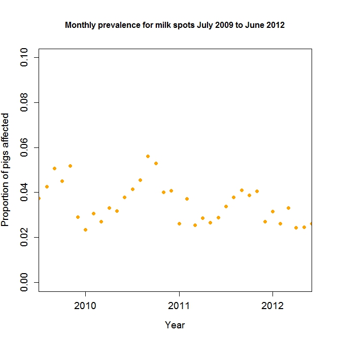
Fig 4 Courtesy of BPHS
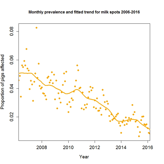
Fig 5 - Courtesy of BPHS
Clinical signs seen with Ascaris infestation will depend on the level of contamination and the site of the larvae or adult.
In the mild cases most commonly seen, the only evidence of the worms is at slaughter where white specks are seen on the liver, giving the term "milk spot". The liver is condemned as a result.
Migration through the lungs can present as coughing in growing pigs - impossible to differentiate from enzootic pneumonia and the lungs may contain petechial haemorrhages throughout the tissue at slaughter although this is often grossly obscured by other pathological lesions and slaughter process artifacts.
With very heavy infestation in growing pigs, the young mature worms can block the intestine leading to vomiting, constipation, jaundice, weight loss and death. This is extremely rare.
Milk spot lesions are themselves transient and will resolve after 40 days. Therefore, if there is evidence of liver damage at slaughter, the problem must be occurring in the finishing area. Where liver damage is severe, weight loss, jaundice and death can occur, although more typically there is a reduction in growth of up to 10% and a degeneration in food conversion efficiency of up to 13% in individuals.
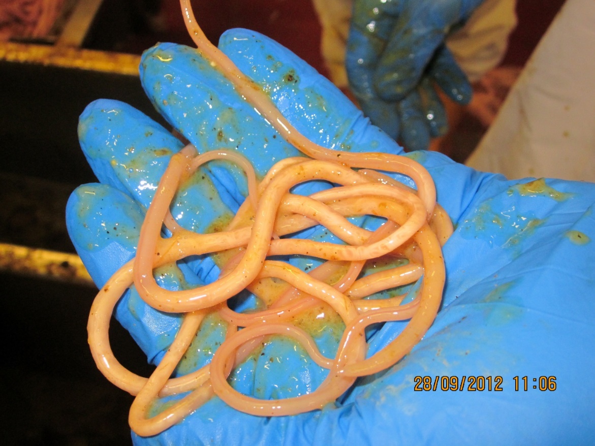
Figure 6 Maturing young adult worms from the gut of a clean pig at slaughter
Within the slaughter house it is also not uncommon to see maturing worms present in the intestines within the gut room. Pigs affected in this way must have been initially infested earlier in life to allow complete migration and the full life cycle. There may well be no or limited milk spot lesions in the liver as these have healed.
Slaughter house monitoring
BPHS reports and to a lesser extent CCIR feedback provide a reliable monitoring system for the levels of parasitism in slaughter pigs. However, lesions need to be differentiated from hepatic scaring which is the result of fibrous tissue build up in the capsule of the liver and is of unknown cause.
Abattoir data collected as part of the Batch Pig Health scheme indicates that up to 90% of herds show no evidence of milk spot livers at slaughter and less than 5% of herds have significant levels of livers affected.
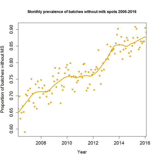
Fig 7 - courtesy of BPHS
In the slaughterhouse batches of pigs fall broadly into four group patterns:
- The vast majority of batches of slaughter pigs show no evidence of milk spot suggesting the husbandry system is sufficient to maintain full control of the parasite or any challenge occurs early in life and lesions have resolved
- Batches containing livers of which many are affected with a small number of lesions suggesting low levels of late challenge either from a low contaminated environment or in a situation where active control of a known problem situation is practiced.
- High levels of contamination in most livers representing a high challenge situation. This is most likely to occur in poor hygiene situations especially where there is continuous occupation and is seen as a particular problem in the organic and free range sectors or where pigs are reared in yards with non-cleanable floor bases.
- The majority of livers are milk spot free but 1 or 2 are heavily infested. Assuming that slapmarks are readable and are accurately read, confirming the anomaly is genuine, the most likely explanation can be that the affected pigs have been grown in a separate environment from the main group. It is highly likely that they will have come through a hospital area which is never cleaned or emptied and contains compromised animals that are most likely to be affected with parasites .
It should also be noted that there are other related non-pig worm parasites which in specific circumstances can spill over into pigs. The most likely scenario is where cats contaminate growing pig environments and the cat roundworm -Toxocara cati - sheds eggs that can produce larvae to be picked up by the pigs. These will migrate to the liver inducing milk spot like lesions but generally do not complete the life cycle.
It should also be noted that A suum being related to the dog and cat roundworms (Toxocara species) have the potential to act as a zoonosis and infect man. Migrating larvae in theory can cause similar problems to these pet parasites (blindness etc) although it is dubious as to whether this has ever been definitively identified.
Control
Where Ascaris has been demonstrated as a significant problem, (for instance more than 25% livers condemned at slaughter) a rigorous cleaning programme is needed to reduce the levels of environmental contamination. Use of a detergent in the cleaning will help to break down the sticky coat of the egg, probably allowing eggs to be washed away. Conventional disinfection is unlikely to have must effect. Final treatment of a washed area with a flame gun is effective, allowing for the obvious health and safety concerns.
Worming of adult sows is advisable to stop further production of worm eggs, although alone is inadequate to control an established problem where environmental contamination is most significant.
Where treatment of growing pigs proves necessary, it is important that the product chosen is effective against the larvae as well as the adults. Such products include the Avermectins (Ivomec, Dectomax) and the benzimidazole group including fenben, (Panacur:Intervet) and Flubendazole (Flubenol: Janssen). Obviously, care must be taken over withdrawal periods - particularly the injectable products. The Avermectins are generally only justifiable on cost grounds if there is a need to control sarcoptic mange as well as worm burdens.
Abattoir data collected as part of the Batch Pig Health scheme indicates that approximately 70% of herds show no significant evidence of milk spot livers at slaughter and only 5-8% of herds have an incidence of more than 25% livers affected.
porternoestringthe1991.blogspot.com
Source: https://www.nadis.org.uk/disease-a-z/pigs/ascariasis/
0 Response to "White Spots in Beef Liver Safe to Eat"
Postar um comentário